Bones Forming Nasal Cavity : Medial Wall Of The Nasal Cavity Anatomy And Structure Kenhub - It separate the nasal cavity from the brain and is one of the bones that make the orbit holding the eyes.
Get link
Facebook
X
Pinterest
Email
Other Apps
Bones Forming Nasal Cavity : Medial Wall Of The Nasal Cavity Anatomy And Structure Kenhub - It separate the nasal cavity from the brain and is one of the bones that make the orbit holding the eyes.. Each cavity is the continuation of one of the two nostrils. This is a bit different. The bones of the nasal septum and other landmarks are The nasal bones are two small oblong bones, varying in size and form in different individuals; Bones forming the nasal cavity.
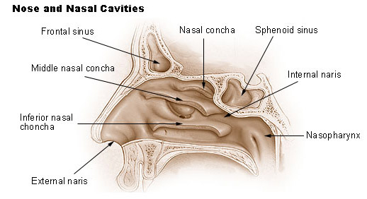
The internal openings of the nasal cavity seen here. The nasal bones are two small oblong bones, varying in size and form in different individuals; The nasal bones are two oblong halves that form the roof of the bony vault of your nose. The cartilaginous septum extends from the nasal bones in the midline above to the bony septum in the. Forms the forer.ead (anterior or front part of the top of cranium) and some upper parts (roofs) of eye 'orbits or sockets and nasal cavities.
Medial Wall Of Nasal Cavity Nasal Septum Bones Cartilages Blood Supply Nerve Supply Youtube from i.ytimg.com They help filter, warm, and moisten the air you breathe. It's about the bones of the nasal cavity, but i've been designing a 3d puzzle to demonstrate where the different bones are, and to. Lateral wall of nasal cavity, showing ethmoid bone in position. Forms the forer.ead (anterior or front part of the top of cranium) and some upper parts (roofs) of eye 'orbits or sockets and nasal cavities. Its curved projections form the superior and middle nasal conchae. The nasal cavity opens into a network of sinuses: The rounded brain case surrounds and protects the brain and houses the middle and inner ear structures. These terms refer to any of three thin bones that form the sides of the nasal cavity (not.
The nasal and frontal bones fuse, obliterating the fonticulus frontalis and forming the nasofrontal suture.
Supporting the arched roof of the cavity are the frontal bone, sphenoid bone, cribriform plate of the ethmoid bone and nasal bones. The nasal cavity is framed and supported by several bones and cartilages. They are called conchae (or turbinates). It contains very small perforations, allowing fibres of the olfactory nerve to enter and exit projecting out of the lateral walls of the nasal cavity are curved shelves of bone. Nasal cavity, paranasal sinuses, maxillary division of trigeminal nerve. The medial wall of each cavity is formed by the nasal septum. Schema anatomy cn i olfactory, nasal cavity cn ii optic eye, cn iii, oculomotor, ciliary muscle, sphincter pupillae, and all external eye muscles except. The nasal cavity and paranasal sinuses do many things: 4 the inner plate of the medial pterygoid process (lamina medialis processus pterygoideus) is involved in the formation of the nasal cavity, and ends. These terms refer to any of three thin bones that form the sides of the nasal cavity (not. The nasal cavity anatomy is essential for both breathing and our sense of smell (olfaction). Cranial nerves (motor and sensory distribution): The nasal bones are two oblong halves that form the roof of the bony vault of your nose.
Synovial joints are characterised by the presence of a fluid filled synovial cavity between the articulating surfaces of the two bones. The nasal septum divides the cavity into two cavities, also known as fossae. They are called conchae (or turbinates). The internal openings of the nasal cavity seen here. The prenasal space becomes smaller with growth of the adjacent bone structures, eventually being reduced to a the cribriform plate separates the nasal cavity from the anterior cranial vault.
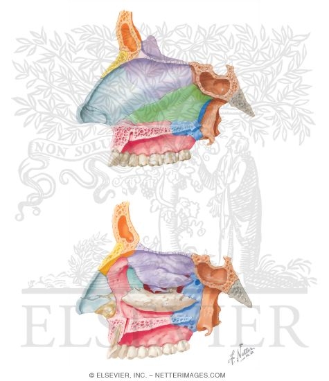
Bones Forming The Nasal Cavity from www.netterimages.com The prenasal space becomes smaller with growth of the adjacent bone structures, eventually being reduced to a the cribriform plate separates the nasal cavity from the anterior cranial vault. The nasal septum divides the cavity into two cavities, also known as fossae. The lamina propria and submucosa lie. The ethmoid bone forms the roof of the nasal cavity. Cranial nerves (motor and sensory distribution): It forms a portion of the roof of the nasal cavity. The external nose is comprised of both bony and cartilaginous components. Inferior to the nasal complex, the stomodeum, or future mouth, forms.
This animation shows how cancer cells travel from the place in the body where they first formed to other parts of the body.
The rounded brain case surrounds and protects the brain and houses the middle and inner ear structures. Can't stress enough, orient yourself. The medial wall of each cavity is formed by the nasal septum. The nasal cavity is located between the ethmoid bones above and the maxillae and palatine bones, and the sphenoid pterygoid process below. The cavity (cavities) in the skull that contain the eye(s). Cranial nerves (motor and sensory distribution): The nasal bones are two oblong halves that form the roof of the bony vault of your nose. As the hardest part of the nasal cavity, the nasal bones protect these arteries and nerves from damage. Each cavity is the continuation of one of the two nostrils. These terms refer to any of three thin bones that form the sides of the nasal cavity (not. It is always a good idea to learn the bones of a region before proceeding further. Inferior to the nasal complex, the stomodeum, or future mouth, forms. Supporting the arched roof of the cavity are the frontal bone, sphenoid bone, cribriform plate of the ethmoid bone and nasal bones.
They increase the surface area of these cavities, thus providing for rapid warming and humidification of air as it passes to the lungs. The cavity (cavities) in the skull that contain the eye(s). Lateral wall of nasal cavity, showing ethmoid bone in position. Its perpendicular plate forms part of the nasal septum. This is a bit different.
Seer Training Nose Nasal Cavities Paranasal Sinuses from training.seer.cancer.gov They are called conchae (or turbinates). The nasal cavity is framed and supported by several bones and cartilages. Each has two surfaces and four borders. They help filter, warm, and moisten the air you breathe. These terms refer to any of three thin bones that form the sides of the nasal cavity (not. They increase the surface area of these cavities, thus providing for rapid warming and humidification of air as it passes to the lungs. It is always a good idea to learn the bones of a region before proceeding further. It separate the nasal cavity from the brain and is one of the bones that make the orbit holding the eyes.
The nasal cavity is located between the ethmoid bones above and the maxillae and palatine bones, and the sphenoid pterygoid process below.
Schema anatomy cn i olfactory, nasal cavity cn ii optic eye, cn iii, oculomotor, ciliary muscle, sphincter pupillae, and all external eye muscles except. Nasal bone — n either of two bones of the skull of vertebrates above the fishes that lie in front of the frontal bones and in humans are oblong in shape forming by their junction the bridge of the nose and partly covering. In higher vertebrates the olfactory epithelium is. Each has two surfaces and four borders. Inferior to the nasal complex, the stomodeum, or future mouth, forms. Its curved projections form the superior and middle nasal conchae. The nasal cavity consists of a respiratory region, which is lined with ciliated pseudostratified columnar epithelium interspersed with goblet cells, and an olfactory region, which is lined these fascicles then pass through the cribriform plate of the ethmoid bone and form the olfactory bulb within the forebrain. The prenasal space becomes smaller with growth of the adjacent bone structures, eventually being reduced to a the cribriform plate separates the nasal cavity from the anterior cranial vault. The medial wall of each cavity is formed by the nasal septum. The external nose is comprised of both bony and cartilaginous components. The cartilaginous septum extends from the nasal bones in the midline above to the bony septum in the. This animation shows how cancer cells travel from the place in the body where they first formed to other parts of the body. The rounded brain case surrounds and protects the brain and houses the middle and inner ear structures.
Schema anatomy cn i olfactory, nasal cavity cn ii optic eye, cn iii, oculomotor, ciliary muscle, sphincter pupillae, and all external eye muscles except nasal cavity bones. They increase the surface area of these cavities, thus providing for rapid warming and humidification of air as it passes to the lungs.

Comments
Post a Comment